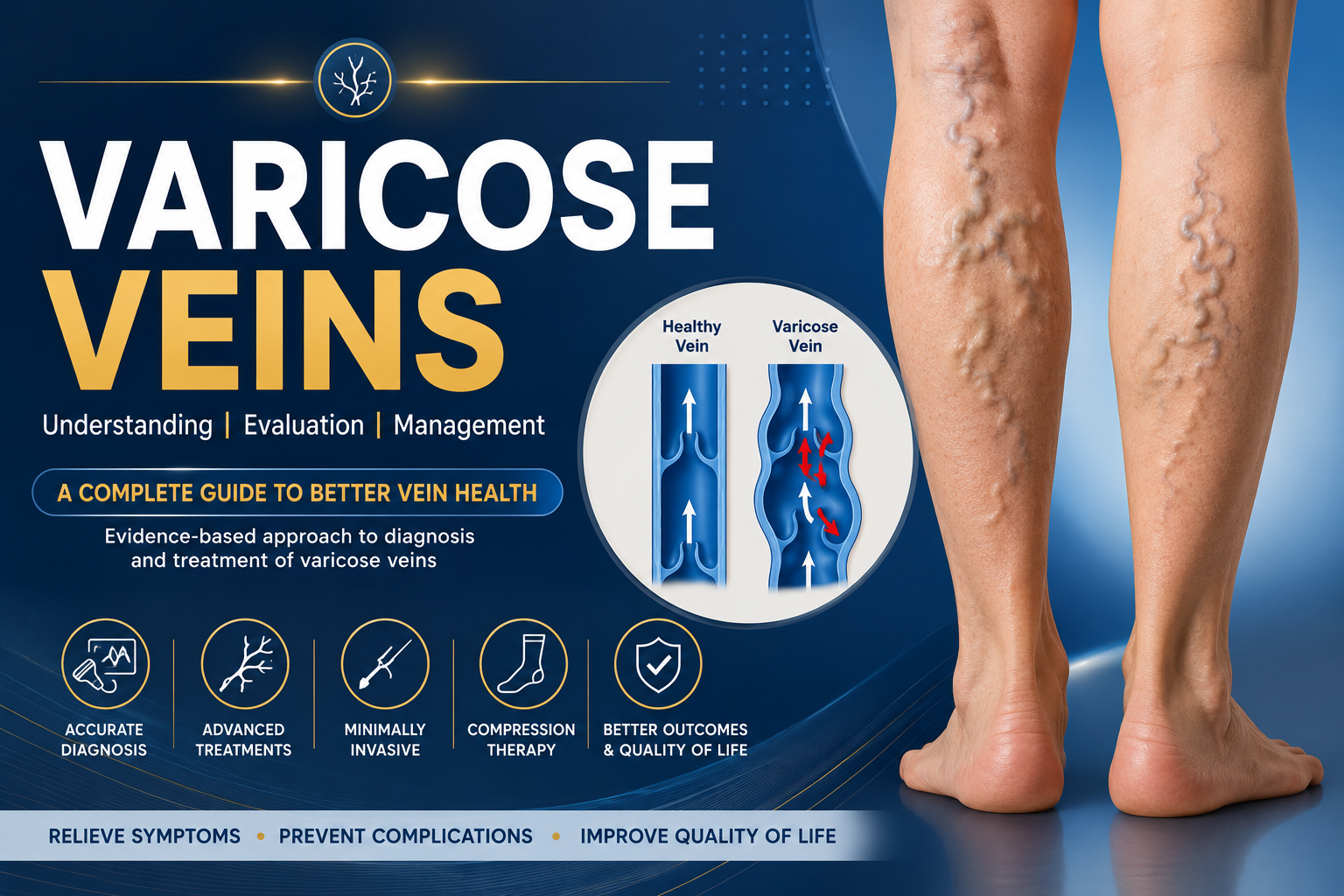
Varicose Veins
Varicose veins are dilated, tortuous superficial veins of the lower extremities, typically ≥3 mm in diameter when measured upright. They result from chronic venous insufficiency (venous reflux and hypertension) in the great saphenous, small saphenous, or perforator veins. Globally, varicose veins affect roughly 10–30% of adults (higher in women than men). Prevalence rises with age, obesity, and pregnancy. Patients often present with leg heaviness, aching, edema, itching or stasis dermatitis, which worsen on standing. Left untreated, varicose veins can cause complications like bleeding, thrombophlebitis, skin changes, and venous ulcers.
Diagnosis is by history and physical exam (including Trendelenburg or Perthes tests) plus duplex ultrasound – the gold standard for mapping venous reflux. The CEAP classification (Clinical–Etiology–Anatomic–Pathophysiologic) grades disease from C0 (no signs) to C6 (active ulcer). Management starts with conservative measures (graduated compression stockings, leg elevation, exercise, weight control) and adjunctive phlebotropic drugs (e.g. diosmin/hesperidin, horse chestnut extract) to relieve symptoms. Definitive treatment of symptomatic truncal reflux usually involves minimally invasive ablation (endovenous laser or radiofrequency, mechanochemical ablation, or cyanoacrylate glue) with or without phlebectomy or sclerotherapy of tributary varices. High-quality trials show endovenous ablation has equal or better efficacy and fewer complications than open surgery. Table 1 (below) compares major treatments by mechanism, efficacy, complications, recovery, and cost. A venous specialist should tailor therapy to patient anatomy, symptoms and comorbidities. Key patient education includes lifestyle modification (avoid prolonged standing, weight management, skin care) and adherence to compression. Recurrence occurs in up to 20–50% by 5–10 years, underscoring the need for follow-up. Current research is focusing on optimizing long-term outcomes of new therapies, cost-effectiveness, and preventive strategies.
Definition and Epidemiology
Varicose veins are defined as permanently dilated subcutaneous veins (typically ≥3 mm in diameter when standing) in the lower limbs. They lie above the muscular fascia and usually involve the great or small saphenous systems and their tributaries. Varicosities can occur in isolation or as part of Chronic Venous Disease (CVD), which includes telangiectasia, edema, skin changes, and ulcers. The CEAP clinical classes are:
- C0: no visible signs
- C1: telangiectasias/spider veins (<1 mm) or reticular veins (1–3 mm)
- C2: varicose veins (≥3 mm)
- C3: edema (without skin changes)
- C4a: skin pigmentation or eczema; C4b: lipodermatosclerosis or atrophie blanche
- C5: healed venous ulcer; C6: active venous ulcer.
In addition to the clinical grade, CEAP classifies etiology (primary, secondary [post-thrombotic or obstruction], or no venous cause), anatomy (superficial, deep, perforator), and pathophysiology (reflux, obstruction, or both). Varicose veins fall under “primary” reflux pathology unless a precipitant (e.g. DVT) is identified.
Globally, varicose veins are very common. Estimates vary widely by study, but approximately 10–30% of adults are affected. Prevalence increases with age and is several times higher in women. In one meta-analysis of healthcare workers, 25% had varicose veins. In the general population, 1–17% of men and up to 40% of women have chronic venous insufficiency, and varicosities have been reported from <1% to 70% (depending on diagnostic criteria). Risk factors include advancing age, female gender, family history, obesity, pregnancy, prolonged standing or sedentary work, prior leg injury or DVT, and hormonal factors (e.g. OCP use). Industrialized nations tend to have higher prevalence due to lifestyle factors.
Complications of chronic venous hypertension develop in more severe disease (CEAP C4–C6). Venous stasis leads to capillary leakage, hemosiderin deposition (brown pigmentation), lipodermatosclerosis and ulceration. About 1–3% of adults develop a venous leg ulcer (CEAP C6) at some point. Even without ulceration, varicose veins can bleed or thrombose. Left untreated, chronic venous disease impairs quality of life and incurs substantial healthcare costs.
Pathophysiology and Etiology
Venous return from the legs depends on competent bicuspid valves in the superficial, deep, and perforator veins and adequate calf muscle pump function. When valves fail or flow is obstructed, venous reflux and hypertension result. Superficial venous reflux is often idiopathic (primary) – due to valve cusp distortion or vein wall weakness (from genetic, inflammatory, or biochemical changes). Over time, increased intraluminal pressure causes veins to dilate and become tortuous (varicose). Most reflux originates at the saphenofemoral junction of the great saphenous vein (GSV) or the saphenopopliteal junction of the small saphenous vein (SSV). Incompetent perforator veins can also transmit high pressure into the superficial system.
Secondary varicose veins arise from deep venous obstruction or post-thrombotic damage. Prior deep vein thrombosis (DVT) scars and narrows veins (post-thrombotic syndrome), causing residual obstruction and reflux. Nonthrombotic iliac vein compression (May-Thurner syndrome) is another example of deep venous outflow obstruction. Rarely, arteriovenous malformations or syndromes like Klippel–Trenaunay–Weber cause limb vein dilation and varicosities. In summary, venous hypertension – whether from valvular reflux or outflow block – is the final common pathway. Persistent high venous pressure leads to capillary damage, edema, skin hyperpigmentation, dermatitis, and ultimately ulceration.
The underlying histology in primary varicose veins often shows vein wall remodeling: increased collagen, reduced elastin, and inflammatory infiltrates that degrade valve structure. Secondary CVI after DVT involves inflammation, vein wall fibrosis and adhesion. Genetic factors (familial tendency) play a role, though specific genes are still being studied. Whether superficial or deep, chronic venous insufficiency perpetuates itself by worsening hypertension and vessel changes.
Risk Factors
Multiple risk factors for varicose veins have been identified. Non-modifiable: female sex (possibly hormonal or obstetric factors), family history of venous disease, taller height, and older age all increase risk. In the StatPearls review, age ≥55 was highlighted. Certain life stages – especially pregnancy – greatly increase risk due to hormonal changes and weight of the gravid uterus. Other non-modifiable factors include genetic syndromes (Klippel–Trenaunay, Ehlers-Danlos) affecting vessel integrity.
Modifiable: Obesity (increased intra-abdominal pressure), prolonged standing or sitting (occupations like nursing, retail), and a sedentary lifestyle reduce calf pump efficiency. Chronic constipation or any cause of elevated intra-abdominal pressure can worsen venous return. History of leg injury or trauma is sometimes associated. A past DVT is a strong risk for secondary varicosities. Smoking has also been cited as a risk. In short, anything that injures valves, increases intra-abdominal pressure, or impairs calf muscle pump can contribute to varicose vein development.
Clinical Presentation and Complications
Patients with varicose veins typically present with leg symptoms and visible vein changes. Early on (CEAP C1–C2), patients may notice spider or reticular veins (thin dilated networks) and larger bulging veins on the calf or thigh. Common subjective symptoms include leg heaviness, aching, pressure, cramping, burning, and itching, especially after prolonged standing. Symptoms often improve with leg elevation or walking. Physical exam may reveal dilated superficial veins, swelling (edema), skin redness or warmth from stasis dermatitis, and a “woody” feel of the skin (lipodermatosclerosis).
As disease advances (C3–C6), chronic venous insufficiency findings appear: persistent edema, venous stasis pigmentation, eczema, and eventually venous ulcers (usually near the medial malleolus). About 1–3% of adults develop venous ulcers in their lifetime. Other complications: superficial thrombophlebitis (tender cord of inflamed vein), bleeding from an exposed varix, and recurrent infections in stasis skin changes. Rarely, large superficial vein aneurysms (focal dilations) can develop and may thrombose or embolize. Patients may also have concomitant deep venous disease (history of DVT, chronic edema) that exacerbates symptoms.
Importantly, varicose veins are often asymptomatic aside from cosmetic concern; treatment decisions hinge on symptoms (pain, swelling, ulceration) and complication risk. Any leg ulcer in a patient with varicose veins should prompt vascular evaluation, as treating underlying reflux improves healing.
Diagnostic Approach
Evaluation of varicose veins starts with a thorough history and physical. Key history: duration of symptoms, family history of venous disease, past leg DVT or injuries, pregnancy history, occupations requiring standing. Ask about claudication to rule out peripheral arterial disease (PAD). On exam: inspect legs in standing position for varices, edema, skin changes (pigmentation, eczema, ulcer). Palpate pulses to exclude arterial disease. Percussion tests (Trendelenburg/Perthes) can help localize reflux to superficial versus perforator veins. For example, in the Trendelenburg test, a rapid fill of veins when a tourniquet is released suggests isolated superficial reflux.
Duplex ultrasound is the gold-standard diagnostic test. It confirms reflux (retrograde flow >0.5 seconds) in superficial, deep, or perforating veins and maps their anatomy. Ultrasound should be performed with the patient upright or using a reverse Trendelenburg position to maximize gravity on venous flow. Often, the protocol examines the saphenofemoral and saphenopopliteal junctions, GSV/SSV course, and perforators. Duplex also evaluates for deep vein patency (prior DVT scars) and measures calf pumping. Sometimes noninvasive tests like air-plethysmography or photoplethysmography are used in research but are not routine. Venography or MR venography is rarely needed except for pelvic or deep obstruction workup.
Classification by CEAP is useful: it requires documenting clinical class (as above), etiology (primary vs secondary), anatomic location (GSV vs SSV vs tributaries), and pathophysiology (reflux vs obstruction) based on imaging. This standardized scheme guides management decisions and comparisons in studies. For instance, CEAP C2S (primary, superficial reflux) indicates simple varicosities without advanced changes.
Differential diagnosis includes pelvic congestion syndrome (in women), lymphedema, lipedema, chronic leg edema from heart/liver/kidney disease, and other causes of leg ulcers (arterial, neuropathic, vasculitic). In practice, proper venous history and duplex usually confirm the cause.
Staging/Classification (CEAP)
The Clinical-Etiology-Anatomic-Pathophysiology (CEAP) classification is the standard staging system for chronic venous disorders. In practice, it is most often summarized by the “C” class (C0–C6) as described under Definition. Primary etiology (E=C) denotes unknown cause (usually valvular reflux) and Secondary (E=CV or CP) denotes post-thrombotic or obstruction. Anatomic (A) codes which veins are affected (e.g. superficial [As], deep [Ad], perforators [Ap]). Pathophysiology (P) denotes reflux (Pr), obstruction (Po), or no venous cause. For example, “C2, Ep, As, Pr” means clinical varicose veins, primary etiology, superficial veins, with reflux. CEAP is useful for research and documenting extent; in clinical summaries one typically mentions CEAP class and the specific veins (e.g. “C4a varicosities with primary GSV reflux”).
Conservative Management
For most patients, conservative (nonprocedural) therapy is first-line. This includes compression therapy, leg elevation, exercise and weight control, and pharmacologic venotonics.
-
Compression stockings: Graduated elastic stockings (Class II, ~30–40 mmHg at ankle) are the mainstay. Compression improves venous reflux, reduces pain/edema, and can even help heal venous ulcers. Stockings should be well-fitted and worn daily (especially during waking hours). Compliance is crucial: benefits occur only if worn regularly. (Caution in severe PAD; obtain ABI first.)
-
Leg elevation and exercise: Elevating the legs above heart level (e.g. lying with legs propped up) for ~30 minutes several times a day reduces venous pressure and swelling. Regular exercise (walking, calf raises, cycling) activates the calf muscle pump, aiding venous return. Ankle flexion/extension exercises help open and close valves, improving flow. Avoid long periods of standing or sitting; take frequent breaks to move legs.
-
Lifestyle: Weight reduction is advised (obesity is a strong risk factor). Dietary sodium reduction can lessen edema. Advise loose clothing and avoiding high heels, which impair calf pump. In pregnancy, compression and avoidance of constipation are recommended. Smoking cessation is generally advised (it impairs circulation).
-
Pharmacotherapy: No drug cures varicose veins, but venoactive drugs (phlebotonics) can relieve symptoms. Agents like micronized purified flavonoid fraction (diosmin/hesperidin), horse chestnut seed extract (Aescin), rutosides, oxerutins or pentoxifylline have modest benefit on symptoms and edema. Evidence quality is limited. One Cochrane found horse chestnut extract reduces leg pain/swelling but long-term safety/efficacy data are sparse. Pentoxifylline (a rheologic agent) can hasten venous ulcer healing (often used with compression). Venoactives are often safe but may cause GI upset. They are adjuncts, not substitutes for definitive therapy.
-
Skin Care: In patients with stasis dermatitis or healed ulcers, moisturization and preventing skin breakdown is key. Keep skin supple (apply lanolin-based creams). For eczematous dermatitis, topical steroids may be used. Foot and leg skin should be inspected daily for abrasions to avoid infection, especially in venous ulcer history.
Compression and lifestyle measures do not eliminate varicose veins but can control symptoms and prevent progression or ulceration. All patients should receive education on these measures, even if proceeding to procedural treatment. Guidelines support early referral to interventional therapy for symptomatic reflux rather than prolonged trials of conservative care, especially when quality of life is affected.
Procedural and Surgical Treatments
When symptoms are significant or complications (ulcer, bleeding) occur, interventional therapy is indicated. Modern treatment has largely shifted to minimally invasive endovenous techniques; traditional surgery is now reserved for select cases. The main options are:
-
Foam sclerotherapy: Injection of a sclerosant foam (e.g. polidocanol, sodium tetradecyl sulfate) into the varicose vein causes endothelial damage and closure. Foam sclerotherapy can be done under ultrasound guidance for larger veins, and with bare-eye (“liquid sclerotherapy”) for small telangiectasias or reticular veins. It is effective for many secondary tributaries and incompetent perforators, often used adjunctively. Drawbacks: multiple sessions may be needed; there is risk of hyperpigmentation, matting, or thrombophlebitis.
-
Endovenous thermal ablation: This is now the first-line treatment for truncal saphenous reflux. It includes:
- Radiofrequency ablation (RFA): A catheter delivers thermal energy (often 120–140 °C) to the vein wall, causing collagen contraction and closure. Usually done under tumescent local anesthesia to minimize pain. RFA occludes the GSV/SSV with ~80–95% success at 2+ years.
- Endovenous laser ablation (EVLA): A laser fiber (usually 810–1470 nm wavelength) is pulled through the vein, causing photothermal destruction. EVLA has similarly high occlusion rates (85–93% at 1–2 years). Some studies suggest EVLA causes more post-procedure pain than RFA, but outcomes are comparable.
Both methods have largely replaced open stripping in many practices. Expected recovery is quick: most patients resume normal activities in 1–3 days. Potential complications: deep vein thrombosis (rare), skin burns (very rare with tumescent technique), nerve injury (especially tibial nerve with SSV treatment), bleeding or infection (very rare).
-
Cyanoacrylate glue (n-butyl-2-cyanoacrylate): A newer non-thermal ablation. Glue is injected endovenously to glue the vein shut. It obviates need for tumescent anesthesia, so is often less painful. Early results show >90% closure rates at 1–2 years. Possible complications: phlebitis along treated vein (usually mild), and theoretical embolism risk if placed too proximally.
-
Mechanochemical ablation (MOCA): Combines mechanical injury (a rotating wire) with liquid sclerosant. The Clarivein system is most common. It can occlude the vein without heat, so no tumescent needed. Early success rates (~90% at 1 year) are similar to thermal ablation. Some evidence suggests a slightly lower efficacy than EVLA/RFA, but fewer anesthetic needs. A meta-analysis found MOCA anatomic success ≈93% at 1 year with very low serious complications. (We cite SCIENCE from [61] hint: “93.2% success, only DVT.”)
-
Ambulatory phlebectomy: Microscopic surgical removal of varicosities through tiny incisions. Under local anesthesia, forceps are used to extract twisted veins. This is ideal for bulging surface veins not treated by reflux ablation. Phlebectomy yields excellent cosmetic results. It can be done concurrently with truncal ablation or staged. Risks: bruising, small scars, rarely nerve irritation. Recovery is very quick (most return to normal same day or next day).
-
Ligation and stripping: Traditional open surgery involves high ligation of the saphenofemoral junction and stripping (removal) of the GSV (sometimes also SSV). This was once standard but is now less common. It requires general or spinal anesthesia and has more downtime (weeks). Guidelines note endovenous ablation may have equivalent or better outcomes with fewer complications than stripping. Indications for stripping now are limited: very large tortuous veins, failed prior ablations, or when endovenous modalities are contraindicated. Ligation/stripping can improve ulcer healing and symptoms, but recurrence rates tend to be higher than with endovenous methods. Complications include wound infection, nerve injury (saphenous/sural), deep vein thrombosis, and anesthesia risks.
-
Perforator vein surgery: In cases of isolated incompetent perforators (often in ulcer patients), subfascial endoscopic perforator surgery (SEPS) or ultrasound-guided perforator ablation (sclero or thermal) can be done. These are specialized interventions.
-
Deep venous interventions: Although beyond “varicose veins” per se, chronic deep venous obstructions (e.g. iliofemoral stenosis) require venous stenting or bypass for ulceration management. Endovascular iliac stenting has largely replaced open venous bypass for May-Thurner syndrome, with 5-year ulcer-free rates ~85–90%.
For varicose tributaries (reticular veins, phlebitic veins), guidelines advise treating concurrently or immediately after treating the truncal reflux. Either phlebectomy or ultrasound-guided foam sclerotherapy is appropriate. The choice depends on vein size and patient preference. Small telangiectasias (<1 mm) are best managed by cosmetic sclerotherapy or laser.
Table 1 below summarizes the mechanism, efficacy, complications, recovery, and cost range of major treatments:
| Treatment | Mechanism | Efficacy (vein closure/symptom relief) | Notable Complications | Recovery Time | Cost (typical U.S. estimate) |
|---|---|---|---|---|---|
| Endovenous RFA | Thermal (radiofrequency ablation of vein wall) | High (≈85–95% occlusion at 1–2 years; significant symptom relief) | Rare DVT/PE; nerve injury (sural nerve in SSV tx); bruise, phlebitis | Outpatient; resume normal in 1–3 days | ~$1,500–$3,000 per leg |
| Endovenous EVLA | Thermal (laser ablation) | High (≈85–93% occlusion); similar to RFA | Similar to RFA (pain may be slightly higher) | 1–3 days | ~$1,500–$3,000 per leg |
| Cyanoacrylate glue | Chemical occlusion (glue) | High (90–95% in short-term) | Phlebitis (inflammatory), rare glue migration | 1–3 days | ~$1,500–$3,000 per leg (varies) |
| MOCA (Clarivein) | Mechanochemical (rotating wire + sclerosant) | ≈90–93% at 1 year (per limited studies) | Minimal; possible DVT (very low) | 1–3 days (no anesthesia) | ~$1,500–$3,000 per leg (est.) |
| Foam sclerotherapy | Chemical (inject sclerosant foam) | Moderate: occludes many tributaries (~70–85% short-term) | Skin staining, matting, superficial thrombophlebitis | Outpatient; immediate return | ~$300–$600 per session |
| Phlebectomy | Ambulatory micro-incision removal | High for removed veins; symptomatic relief good | Bruising, small scars, nerve irritations (rare) | 1–2 days (local anesthesia) | ~$2,000–$5,000 per leg |
| Ligation/Stripping | Surgical removal of vein | High initial, but recurrence higher than EVLA | Infection, nerve injury, DVT, hematoma | Weeks (requires anesthesia) | Unspecified (higher than endovenous) |
Table 1. Comparison of varicose vein treatments. Efficacy and complications are approximate; actual outcomes depend on patient factors. Cost ranges from U.S. sources (e.g. specialist centers) for single-leg treatment.
Comparative Effectiveness
Multiple studies and meta-analyses have compared varicose vein treatments. Overall, endovenous ablation (EVLA/RFA) is as effective as open surgery for vein closure, with less postoperative pain and faster recovery. A Cochrane review found no significant difference in occlusion rates between RFA and EVLA, but RFA had fewer post-op complications. For superficial veins, EVLA/RFA achieve >80% occlusion at 1–2 years, whereas stripping or phlebectomy also have high short-term success but higher long-term recurrence due to neovascularization.
Comparing EVLA vs foam sclerotherapy, EVLA is generally more effective at occluding large truncal veins. One randomized trial showed EVLA was superior to ultrasound-guided foam sclerotherapy (UGFS) for GSV closure at 12 months, with better quality-of-life scores. Foam is typically reserved for smaller veins or adjuncts. MOCA vs RFA/EVLA: limited data suggest similar 1-year occlusion (around 90%); MOCA’s main advantage is avoiding tumescent anesthesia. Glue vs RFA/EVLA: early results are promising (~95% success at 1 year) but long-term data are pending.
For varicose tributaries, a randomized trial found no difference in cosmetic outcome between phlebectomy and liquid sclerotherapy when combined with stripping. Thus, choice can be based on patient factors. In summary, minimally invasive ablation has become the standard for truncal reflux, with equivalent or better outcomes than surgery. Combination therapy (ablation + adjunctive treatment) often provides the best symptomatic relief and appearance.
Indications and Contraindications
Indications: Interventions are indicated for symptomatic varicose veins (leg pain, swelling, heaviness) when conservative measures fail, or for complications (ulceration, bleeding). Reflux must be documented (typically by duplex). Active or healed venous ulcers (CEAP C5–C6) with superficial reflux are also indications for venous intervention (it promotes ulcer healing). Cosmetic-only reasons are less justifiable unless the varicose veins cause significant discomfort or risks. Pregnancy: Treatments are generally postponed until after delivery unless complications demand it (e.g. active ulcer).
Contraindications: Acute deep vein thrombosis is a contraindication to treat varicosities (address DVT first). Severe peripheral arterial disease (ABI <0.5) is a contraindication to compression therapy. Class III–IV heart failure can limit ability to tolerate compression or surgery. In endovenous procedures, local infection at insertion site, coagulopathy, or allergy to sclerosant/adhesives are cautions. For RFA/EVLA, inability to lie still or cooperate is a practical barrier. All procedures should be avoided in patients with life expectancy or mobility so poor that quality-of-life gains are minimal.
Perioperative Care
Most varicose vein interventions are outpatient or ambulatory procedures. Preparation includes mapping veins with ultrasound (even on day of procedure). For endovenous thermal ablation, tumescent anesthesia (large-volume dilute lidocaine around vein) is placed under ultrasound. Patients may receive aspirin perioperatively (low-dose) if no contraindication, though routine DVT prophylaxis (e.g. heparin) is usually not required for isolated superficial procedures unless other risk factors exist. Graduated compression stockings (Class II) are typically applied immediately post-procedure.
After surgery or ablation, early ambulation is encouraged (walking the same day). Patients should wear compression stockings continuously for ~1–2 weeks (or as directed) to reduce bruising and edema. Ice packs and NSAIDs help pain/swelling. Weight-bearing activities are allowed, but heavy lifting or vigorous exercise should be avoided for a few days. Showering is usually permitted after 24–48 hours; incision sites (if any) kept clean and covered initially. Follow-up visit within a month to check healing and possibly repeat ultrasound is typical. For ulcer patients, continued compression and wound care are vital.
Outcomes and Recurrence
Endovenous ablation success rates are high: studies report venous closure rates around 85–95% at 1–2 years. Quality-of-life and symptom scores improve markedly after ablation or surgery. Complication rates are low in experienced hands. A large registry (the VIRTUS registry) found few major complications for RFA/EVLA (DVT in <1%, nerve injury <2%) with >90% patient satisfaction.
However, varicose veins have a significant long-term recurrence rate. Recanalization of treated vein or development of new reflux in other veins can occur. Reports suggest 20–50% recurrence at 5–10 years after surgery or ablation. Recurrence is often due to neovascularization at the saphenofemoral junction or untreated accessory veins. The more comprehensive the original treatment (e.g. including tributaries), the lower the recurrence. Phlebectomy alone (without treating reflux) almost guarantees recurrence of underlying disease.
Good predictors of durability include complete ablation of reflux and patient adherence to compression. After recurrence, re-treatment (repeat ablation or phlebectomy) can again relieve symptoms. Longitudinal studies show that treating reflux in patients with healed venous ulcers leads to ulcer healing in ~90% of cases; the ulcer recurrence rate is also reduced with venous intervention plus compression.
Cost and Availability
Costs vary by healthcare system and technique. Endovenous procedures are more expensive than conservative care but less morbid than surgery. In the U.S., typical provider charges (non-cosmetic) are roughly: RFA/EVLA $1,500–$3,000 per leg, phlebectomy ~$2,000–$5,000, and single-session sclerotherapy ~$300–$600. Cyanoacrylate and MOCA are similar to RFA. Open surgery (stripping) has historically been more costly due to anesthesia and facility fees. However, many insurers require failure of conservative therapy for coverage.
Global availability differs: in some regions EVLA/RFA are widely used; in others, only stripping/sclerotherapy are available. The shift toward office-based endovenous treatments has made modern therapies more accessible. High-volume vein centers often negotiate costs lower than large hospital OR settings. Cost-effectiveness studies suggest early ablation is cost-effective if it prevents ulcers and enhances quality of life. Economic data in developing countries are limited, but generic sclerosants and compression may be the mainstay where technology is scarce. Emerging low-cost procedures (like MOCA or glue using off-label materials) may broaden access.
Patient Education and Prevention
Patient counseling is essential. Patients should understand that varicose veins are a chronic condition; without addressing reflux, veins will generally worsen over time. Education topics include: the importance of compression stocking compliance, leg elevation when resting, weight control, and avoiding tight clothing. Emphasize skin care (daily inspection, moisturizing) to prevent ulceration. Warn about signs requiring prompt attention: non-healing ulcers, cellulitis (red streaks), or sudden severe pain/swelling (possible DVT).
Preventive measures are similar to conservative therapy: routine exercise, ankle pump exercises, and periodically elevating the legs can delay progression. Pregnancy education: early use of compression can lessen pregnancy-related varicosities. There is no guaranteed prevention, but addressing risk factors (quit smoking, treat constipation, maintain ideal weight) helps.
Patients should be made aware of treatment options and expected outcomes. Shared decision-making is advised: discuss the benefits (symptom relief, ulcer healing) versus risks (nerve injury, DVT) of procedures. Many patients benefit from hearing that modern endovenous treatments are relatively low-risk with quick recovery. Informed patients can also better comply with post-procedure care (stockings, follow-ups).
Research Gaps and Future Directions
Despite advances, several areas need further study. Long-term comparative trials of ablation technologies (e.g. RFA vs EVLA vs glue vs MOCA) are limited; head-to-head 5–10 year data would clarify durability differences. The role of venoactive drugs needs more high-quality RCTs to define which formulations and patient subgroups benefit most. Likewise, better evidence for compression (optimal pressure, duration, and necessity in various CEAP classes) is needed.
Cost-effectiveness analyses in diverse healthcare settings are sparse; given the high cost burden of ulcers, economic models could guide public health policy on varicose treatment. Also, genetic and molecular studies of vein wall biology may identify targets to prevent valve failure.
On the device front, novel adhesives or catheter-based treatments (e.g. glue formulations with faster polymerization) are under development. Telemedicine and remote monitoring for postoperative care (e.g. smartphone Doppler or patient apps) could improve compliance. Finally, since lifestyle is crucial, research into behavioral interventions (digital nudges for exercise or weight loss) could supplement medical care.
In summary, varicose veins remain a common chronic disease. Evidence-based guidelines (e.g. SVS/AVF 2022–2023; ESVS 2022) recommend prompt evaluation and modern therapies for symptomatic reflux. This review highlights current understanding of varicose veins and underscores areas where more data will refine prevention and management.
Mermaid Chart: Treatment Algorithm
Here is a cleaner and professionally formatted version of your flowchart in Mermaid syntax:
Clinical Decision Path
Symptomatic Varicose Veins
│
▼
Active ulcer or severe skin changes?
┌─────────────┴─────────────┐
Yes No
│ │
▼ ▼
Compression therapy Duplex Ultrasound
Wound care │
Consider venous intervention │
┌──────────────┴──────────────┐
Truncal reflux No reflux
│ │
▼ ▼
Endovenous treatment Conservative care
(EVLA / RFA / Glue / MOCA) or Sclerotherapy
│
▼
Phlebectomy or Sclerotherapy
This version follows current evidence-based management pathways for patients presenting with symptomatic varicose veins and is suitable for inclusion in medical articles, clinical guidelines, presentations, or educational materials.